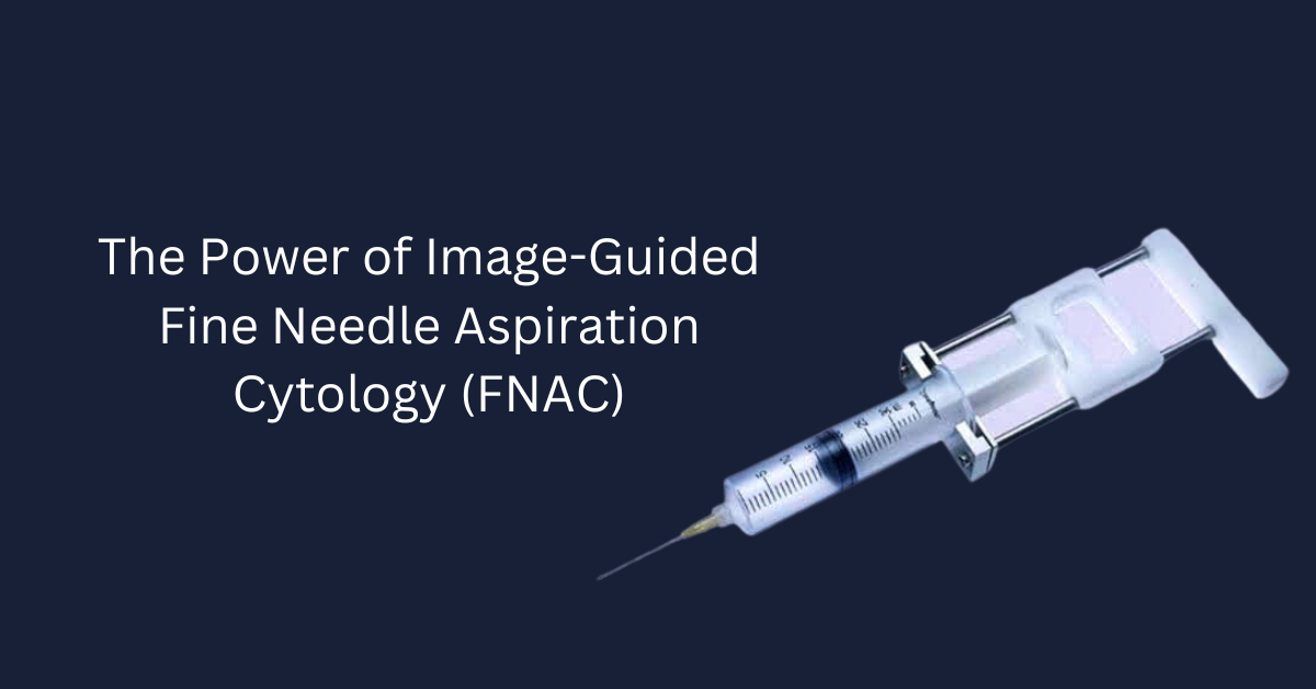
Precision in Diagnosis: The Power of Image-Guided Fine Needle Aspiration Cytology (FNAC)
In the ever-evolving landscape of medical diagnostics, one technique stands out as a true game-changer, offering precise insights into the human body with minimal invasiveness: Image-Guided Fine Needle Aspiration Cytology, or simply FNAC. This remarkable procedure blends cutting-edge imaging technology with the finesse of fine needle aspiration to provide an accurate diagnosis of a wide range of conditions, from benign cysts to malignant tumors. In this article, we will delve into the world of FNAC, exploring how it works, its applications, and the invaluable role it plays in modern medicine.
Table of Contents
The Art of FNAC: A Brief Overview
Fine Needle Aspiration Cytology (FNAC) is a diagnostic procedure that involves the extraction of cells or tissue fragments from a patient’s body using a fine-gauge needle. These extracted cells are then analyzed under a microscope to determine their nature and characteristics. What sets FNAC apart from other diagnostic techniques is the use of real-time imaging guidance, allowing medical professionals to target specific areas with unparalleled precision.
The Process Unveiled: How FNAC Works
Patient Evaluation: FNAC begins with a thorough evaluation of the patient’s medical history, physical condition, and symptoms. This initial step helps medical practitioners determine whether FNAC is the most suitable diagnostic approach.
Imaging Guidance: The defining feature of FNAC is the use of imaging techniques like ultrasound, computed tomography (CT), or fluoroscopy. These imaging methods enable the medical team to visualize the target area, ensuring accurate needle placement.
Needle Aspiration: Once the target is identified, a fine needle, often no thicker than a strand of hair, is inserted into the area of interest. This needle may be guided by the naked eye in some cases but is most effective when paired with real-time imaging guidance.
Sample Collection: As the needle reaches the target, it is used to aspirate a small sample of cells or tissue fragments. This process is relatively painless and minimally invasive.
Sample Processing: The collected sample is then processed and prepared for cytological examination. This involves creating slides and staining the cells for microscopic analysis.
Cytological Examination: A pathologist examines the stained cells or tissue fragments under a microscope. This examination reveals crucial information about the nature of the cells, whether they are benign, malignant, or indicative of a specific disease.
Diagnosis: The pathologist generates a diagnosis based on their analysis, which is then communicated to the patient’s healthcare team for further action.
Applications of FNAC: A Diagnostic Swiss Army Knife
FNAC is an incredibly versatile diagnostic tool, making it a valuable asset across various medical specialties. Here are some of the primary areas where FNAC shines:
- Oncology
FNAC plays a pivotal role in the diagnosis and staging of cancer. By accurately assessing tumor characteristics, such as its type, grade, and malignancy, oncologists can determine the most appropriate treatment plan for patients. Whether it’s breast, thyroid, lung, or prostate cancer, FNAC can provide crucial insights quickly and with minimal discomfort to the patient.
- Thyroid Disorders
The thyroid gland is a common site for FNAC. It aids in distinguishing between benign thyroid nodules and thyroid cancer, enabling physicians to make informed decisions about surgical intervention or ongoing monitoring.
- Breast Health
Breast FNAC is used to investigate suspicious breast lumps or abnormalities detected during mammography or clinical examination. It helps differentiate between benign conditions, such as fibroadenomas, and breast cancer, guiding treatment choices.
- Abdominal and Pelvic Lesions
In cases of liver, kidney, or ovarian cysts or masses, FNAC is employed to determine their nature. This assists in differentiating between benign and malignant growths, directing subsequent management.
- Lymph Node Assessment
Swollen lymph nodes can be indicative of various conditions, including infections and lymphomas. FNAC of lymph nodes aids in diagnosing the underlying cause, facilitating timely treatment.
- Pulmonary Medicine
FNAC is instrumental in diagnosing lung lesions, including nodules and masses, which could be benign or malignant. Rapid diagnosis allows for prompt intervention in lung cancer cases. Coordinate with a pulmonology specialist regarding this.
Advantages of FNAC
The widespread adoption of FNAC in modern medicine can be attributed to its numerous advantages:
- Minimally Invasive: FNAC is a minimally invasive procedure, which means it involves less pain, shorter recovery times, and reduced risk of complications compared to surgical biopsy methods.
- Real-Time Precision: The ability to use imaging guidance in real time ensures that the needle is accurately positioned, minimizing the chance of sampling errors.
- Speed and Efficiency: FNAC provides rapid results, allowing for timely diagnosis and treatment planning.
- Low Cost: FNAC is often more cost-effective than surgical biopsies and other diagnostic methods, making it accessible to a broader range of patients.
- Reduced Discomfort: Patients generally experience minimal discomfort during and after the procedure.
- Versatility: FNAC can be used to diagnose a wide range of conditions across various medical specialties such as Interventional Radiology Hospital in Coimbatore.
Potential Limitations and Considerations
While FNAC is a highly effective diagnostic tool, it’s essential to acknowledge its limitations:
- Sample Adequacy: Sometimes, FNAC samples may not yield enough material for a conclusive diagnosis, necessitating additional procedures.
- Operator Skill: The accuracy of FNAC largely depends on the skill and experience of the medical professional performing the procedure.
- False Negatives: FNAC may occasionally produce false-negative results, particularly in cases where the sampled tissue is not representative of the entire lesion.
- Risk of Infection: As with any invasive procedure, there is a minimal risk of infection at the needle insertion site.
- Patient Cooperation: Patients must remain still during the procedure to ensure accurate needle placement.
FNAC stands as a beacon of precision, offering timely and accurate insights into a myriad of medical conditions. With its minimally invasive nature, real-time imaging guidance, and versatility across various medical specialties, FNAC has revolutionized the way we diagnose and treat diseases. As technology continues to advance and our understanding of cellular pathology deepens, FNAC will undoubtedly play an even more pivotal role in the future of healthcare, providing patients with quicker, less invasive, and more cost-effective diagnostic solutions.


